IPM Vision Group
![]()
|
IPM Vision Group |
|
publication - software - links - people
 |
Biomedical Person Identification via Iris Recognition |
Two Applications of LPC and Computational Geometry to Computer Vision.
Ali Farhadi, Masoud Alipour, Nima Razavi.
Introduction to human eye and Iris biomedical features
|
In Figure.1 you can see the over all structure of
human eye.
Figure 1. Eye Structure
Human Eye has three distinct layers, each performing a different
task. First interior layer is the retina.
The retina is the most important layer because it
contains visual cells.
Next layer is the choroid which is a vascular membrane
containing large branched pigmented cells. Iris is a part of
choroid . The third layer is the sclera which is the outer coat
which encloses the eyeball except for the front part which is
covered by the cornea.
Conjunctiva : the mucous membrane that lines the inner surface
of the eyelids and is continued over the forepart of the eyeball
.
Macula, also called the yellow spot, is a small yellowish area
lying slightly lateral to the center of the retina that
constitutes the region of maximum visual acuity.
Vitreous is the clear colorless transparent jelly that fills the
eyeball posterior to the lens .
Iris is the opaque contractile diaphragm perforated by the pupil
and forming the colored portion of the eye .
|
|
|
Figure.2 The structure of the iris seen in a transverse section |
The visual properties of human Iris are:
1.crypts. They are hard to
show on this picture. They
are like valleys in the iris.
2. Pigment Spots.
3.Radial and concentric.
furrows, which are the muscles which are used for the expansion or contraction of the iris.
4.collarette separates the ciliary and pupillary areas .
5. Pigment frill .
|
|
Figure 3. The structure of the iris seen in a frontal sector. |
|
| First of all we de-noised our original image by an application of daubechies wavelets. Then we take the edge of the image. We extract iris from the original image by using circular Hough transform. | ||
 original image |
 edge image |
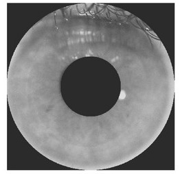 extracted iris |
| In our approach we make use of our feature discrimination and classification
algorithms and some computational geometry approaches.
In the first step after extraction of the iris from the original image, we split our image into several sectors by sweeping our image in polar coordinates with different radiuses and angels. In the second step we compute the LPC coefficients for each sector and put them in a matrix with coordinates of radiuses and angels. The first 9 coefficients of DCT of the matrix (the dc coefficient is discarded) constitutes the first 9 coefficients of our feature vector. The next 3 coefficients of our feature vector are gotten from geometric analysis of LPC matrix. The 13th coefficient of our feature vector is the convex closure of fft analysis of the LPC matrix. To the de-noised image we apply circular DCT, discard the lowest frequency and calculate the kurtosis of each circle. One obtains a set of positive numbers parametrized by the radius of the concentric circles. The kurtosis of this set of numbers is the fourteenth coefficient of the feature vector. For further information click here.
|
| For classification of feature vectors, we clustered our data with a modified k-means clustering algorithm using a weighted distance as our distance function. |
| We have tested our algorithm with our database containing about 400 images of about 70 people (of both eyes) and they are taken over different illuminations and poses. |
Author:
Nima Razavi.
© Copyright 2003-2005 - Institute for Studies in Theoretical Physics
and Mathematics (IPM).
All rights reserved. Please submit your comments or questions
here.
Last update:10/10/2005.


